
25 — Work
Molar root canal treatment
Back teeth can contain curved, divided, hidden, or previously altered canal systems. The plan begins with diagnosis, restorability, and case difficulty—not a promised number of canals or visits.
Original editorial concept · temporary
What it is
A molar is a posterior tooth built to carry substantial chewing load. Its roots and internal canal system are not standardized: canals can curve, divide, merge, flatten into fins and isthmuses, calcify, or sit beneath restorations that obscure the original anatomy. Upper molars commonly require careful evaluation of the mesiobuccal root for additional anatomy; lower molars can contain complex mesial systems and occasional C-shaped or accessory configurations. These population patterns guide a search, but they do not predict one patient’s tooth. Responsible planning establishes the pulpal and apical diagnoses, tests whether the tooth can be isolated and restored, grades the case difficulty, and then selects treatment, referral, imaging, or an alternative. A webpage cannot count the canals, confirm a crack, promise anesthesia, or predict retention.
For
01
Someone told that a first or second molar may need root canal treatment and wants to understand why posterior-tooth anatomy can change difficulty, referral, imaging, and restoration decisions.
02
A patient with pain, biting tenderness, swelling, a deep restoration or crown, calcified or curved canals, an unfinished access, or a previously treated molar that still needs diagnosis.
03
Someone comparing vital-pulp care, nonsurgical root canal treatment, specialist referral, retreatment, apical surgery, extraction with or without replacement, or monitored no treatment.
01 — Diagnose the pulp, root tissues, and pain source
History, examination, sensibility findings, percussion, palpation, periodontal probing, restorability, cracks, adjacent teeth, and appropriate images are interpreted together. Molar pain can be referred, and one symptom or image is not a diagnosis.
02 — Grade difficulty before access
Tooth position, mouth opening, isolation, existing crowns or posts, calcification, curvature, root length, divided or C-shaped anatomy, resorption, nearby structures, prior treatment, and clinician capability influence whether treatment or referral is responsible.
03 — Map anatomy proportionately
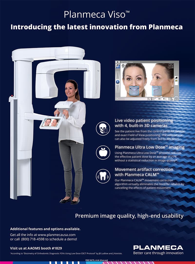
Angled intraoral images and visual anatomic landmarks are foundational. Limited-field CBCT may be justified when a defined complex-anatomy, previous-treatment, resorption, fracture, or nonhealing question cannot be answered adequately with lower-dose information; it is not a routine scan.
04 — Create access that supports treatment
Access preserves sound structure while still permitting canal location, safe instrumentation, irrigation, and obturation. An ultraconservative opening is not automatically safer if it hides anatomy or compromises cleaning; the outline is case-specific.
05 — Disinfect, fill, and protect the tooth
Dental-dam isolation, working-length control, mechanical preparation, irrigation, intracanal medication when indicated, and obturation address the canal system. A sealed interim and definitive coronal restoration are part of the biological and structural plan.
06 — Review the result and retain alternatives
Symptoms, function, restoration, coronal seal, and comparable imaging are reviewed over time. Persistent disease can lead to observation, nonsurgical retreatment, endodontic surgery, extraction, or reconsideration of the diagnosis rather than automatic repetition of one procedure.
Posterior endodontic guide
A molar is not one canal enlarged three times. It is a variable system inside a load-bearing tooth.
The central questions are whether the pulp and apical tissues require treatment, whether the tooth is maintainable, what anatomy and procedural risks are present, who should manage them, and how the restored tooth will be reviewed. Canal counts and technology names come after those decisions.
First and second molars are difficulty signals, not diagnoses
The AAE case-assessment framework classifies a first molar as at least moderate by tooth position and a second or third molar as high difficulty, while the final grade also depends on patient, diagnostic, anatomic, restorative, and previous-treatment factors. The form supports case selection, referral, and documentation; it does not guarantee an outcome or replace clinician judgment.
Internal anatomy varies more than the crown suggests
A root canal system can include main canals, lateral branches, fins, isthmuses, loops, divisions, and apical ramifications. A canal may leave the chamber as one path and divide, or begin separately and merge. Calcification and prior restorations can make the chamber floor and canal path less visible. Therefore, neither a familiar crown shape nor an average canal count is a complete map.
MB2 is a search target, not a promised fourth canal
Systematic reviews using CBCT report that a second mesiobuccal canal is common in maxillary molars, especially first molars, but estimates vary substantially by tooth, population, study method, and region. The safe patient statement is that additional mesiobuccal anatomy should be considered and investigated—not that every upper molar has four canals or that an image alone proves one is negotiable.
Lower molars have their own complexity
Mandibular molars often contain separate mesial and distal systems, while additional middle-mesial canals, root variations, and C-shaped configurations occur in selected teeth and populations. These forms can create narrow fins or communicating spaces that are difficult to instrument directly. Actual anatomy is established for the individual tooth, not assigned from ethnicity or a prevalence table.
Diagnosis and restorability come before canal treatment
The clinician combines symptoms, dental and medical history, sensibility tests with control teeth, percussion, palpation, periodontal probing, mobility, caries and restoration assessment, crack evaluation, and imaging. A vertical root fracture, nonrestorable loss of tooth structure, severe periodontal compromise, or another pain source can change the plan even when the pulp is inflamed or necrotic.
Referral is a quality decision
Restricted opening, difficult anesthesia, severe symptoms or swelling, a second molar, marked inclination or rotation, limited isolation, an altered crown, very long or highly curved roots, calcification, C-shaped or divided anatomy, resorption, proximity to important structures, and previous access or treatment complications all increase difficulty. When the combination exceeds the treating clinician’s competence, equipment, or rescue capacity, consultation or referral is appropriate.
CBCT is selective and question-led
The current AAE/AAOMR position is that CBCT should not be routine. Existing images and the clinical examination come first; a limited field can be considered when three-dimensional information is expected to change diagnosis or management in a complex case. Exposure should be indication- and patient-specific, the field should be as limited as diagnostically acceptable, and the entire acquired volume must be interpreted competently.
Magnification and illumination support—but do not certify—the search
Enhanced illumination, magnification, ultrasonic troughing, careful chamber-floor inspection, and angled radiographs can help locate additional anatomy. They do not prove that every canal exists, can be negotiated safely, or has been completely disinfected. Technology changes the information and control available to a trained operator; it is not an outcome guarantee or proof of local equipment.
The smallest access is not automatically the best access
Preserving pericervical dentin and sound structure is important, but access must still permit safe canal detection, instrumentation, irrigation, and obturation. Systematic-review evidence is largely laboratory-based and low certainty; very contracted access has shown a greater risk of undetected canals when not supported by appropriate magnification and ultrasonic techniques. The practical objective is the smallest access that does not compromise treatment quality.
Dental-dam isolation is a treatment boundary
Isolation separates the operating field from saliva and reduces aspiration or ingestion risk from instruments and irrigants. Existing crown contours, extensive breakdown, or limited remaining structure can make isolation more complex and may require pretreatment modification. Inability to isolate predictably is a reason to revise the plan, not to normalize an unprotected procedure.
Shaping serves irrigation; it is not the biological endpoint
Mechanical instruments create a reproducible path and remove tissue, but they cannot directly contact every fin, isthmus, or irregular surface. Irrigation and, when indicated, intracanal medication address areas instruments do not touch. Curvature and thin root walls require a balance between enlargement, disinfection, transportation risk, perforation risk, and fracture resistance.
Procedural risks must be named without dramatizing them
Case-specific risks can include inability to locate or negotiate anatomy, ledge formation, canal transportation, perforation, separated instruments, irrigant or filling-material extrusion, loss of tooth structure, fracture, persistent symptoms or infection, damage to an existing restoration, need for additional treatment, and tooth loss. A complication does not automatically establish negligence, and its management depends on location, disease, retrieval risk, prognosis, and alternatives.
Missed anatomy is associated with persistent apical disease
A 2025 systematic review found an association between CBCT-detected missed canals and post-treatment apical periodontitis, but the included studies were cross-sectional, heterogeneous, and judged low certainty. That supports careful diagnosis and anatomy review; it does not prove that every persistent lesion is caused by a missed canal or allow one odds ratio to predict an individual tooth.
The restoration is part of the prognosis
Molars bear substantial occlusal load, and access often coexists with caries, cracks, prior fillings, or a crown. The definitive design depends on remaining walls and cusps, crack extent, ferrule, periodontal support, opposing contacts, parafunction, material, and isolation. Cuspal coverage may be appropriate for many structurally compromised molars, but root canal treatment does not create an automatic universal-crown rule and a crown cannot make an unrestorable tooth restorable.
One visit, several visits, and interim care are case decisions
Diagnosis, symptoms, drainage, anatomy, microbial control, procedural events, available time, and restoration logistics can alter sequencing. Evidence does not justify presenting one-visit or multiple-visit care as universally superior. If treatment is staged, the interim seal, temporary-restoration limits, contact route, and conditions for prompt reassessment must be explicit.
A previously treated molar starts with re-diagnosis
Persistent symptoms or apical findings can reflect coronal leakage, untreated or inaccessible anatomy, filling limitations, fracture, periodontal communication, extraradicular factors, scar, or nonendodontic disease. Options can include observation with controls, nonsurgical retreatment, endodontic surgery, extraction with or without replacement, or another diagnostic pathway. Existing posts, cores, crowns, separated instruments, and perforations change access and risk.
Third molars are a separate decision
This guide focuses on permanent first and second molars. A wisdom tooth’s position, access, function, periodontal and caries status, relation to adjacent teeth and vital structures, and long-term maintainability can make extraction or referral considerations materially different. A third molar should not inherit a first-molar treatment assumption from this page.
Follow-up measures healing and restoration together
Review can include symptoms, biting comfort, swelling or sinus tract, percussion and palpation findings, function, periodontal findings, the restoration and coronal seal, and comparable imaging when indicated. Clinical comfort can precede radiographic repair, while absence of pain does not prove biological resolution. No fixed success percentage or healing date should be promised online.
Urgent and emergency boundaries remain explicit
Prompt dental assessment is appropriate for increasing pain, swelling, drainage, fever or malaise, difficulty opening the mouth, a lost interim restoration, fracture, or worsening after treatment. Difficulty breathing or swallowing, rapidly progressive facial or neck swelling, eye symptoms, confusion, collapse, or another life-threatening concern requires 911 or the nearest emergency department.
Continue the evaluation
Clinical reading
General information only. Diagnosis, treatment, risks, alternatives, and expected recovery are individual and require a clinical evaluation.
- ESE S3 guideline: Treatment of pulpal and apical disease ↗
- AAE: Endodontic Case Difficulty Assessment Form ↗
- AAE: Treatment Standards ↗
- AAE/AAOMR: 2025 CBCT position statement release ↗
- PubMed: MB2 prevalence systematic review ↗
- PubMed: Missed canals and post-treatment disease systematic review ↗
- PubMed: Conservative access systematic review ↗
- PubMed: Outcomes of primary root canal therapy systematic review ↗
- Russian peer-reviewed study: Maxillary molar canal curvature ↗
A responsible molar endodontic plan should make seven things explicit: the pulpal and apical diagnoses, restorability, expected anatomy and difficulty, treating-clinician or referral decision, imaging and access rationale, definitive restoration, and the review or rescue pathway.
Request a visit