
13 — Work
Wisdom teeth removal
A decision to remove, monitor, or modify the plan—made from disease, anatomy, risk, and your priorities.
Original editorial concept · temporary
What it is
Third molars may be fully erupted, partly erupted, or impacted. A lack of pain does not prove that a tooth is disease-free; impaction alone does not make surgery automatic. Evaluation connects the medical and dental history, examination, eruption status, periodontal and caries findings, the adjacent second molar, and appropriate imaging. When disease or significant future risk supports surgery, removal and reasonable alternatives are discussed. When neither is present, active clinical and radiographic surveillance is a legitimate plan.
For
01
Pain, swelling, recurrent inflammation or infection, food trapping, decay, or difficulty cleaning around a third molar.
02
An erupted, partially erupted, or impacted tooth with disease—or a meaningful risk to the adjacent molar, supporting tissues, or another planned treatment.
03
A patient with quiet or asymptomatic wisdom teeth who wants a balanced opinion about removal versus active surveillance.
01 — Establish the condition
Symptoms are traced to their source, and the examination records eruption, hygiene access, decay, periodontal health, the adjacent tooth, and any evidence of infection or other pathology.
02 — Image proportionally
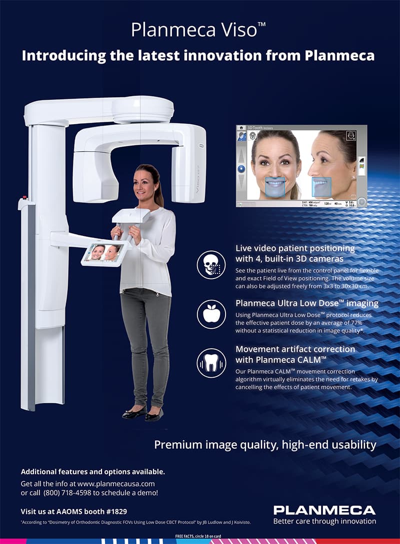
Conventional imaging may be enough. CBCT is reserved for a specific anatomical question that cannot be answered adequately on standard images and could affect management or consent.
03 — Compare the paths
Removal, active surveillance, and—when anatomy makes them relevant—modified surgical approaches or specialist referral are compared without presenting one route as automatic.
04 — Consent, treat, and follow
The agreed plan defines anesthesia, patient-specific surgical risks, recovery expectations, written aftercare, follow-up, and exactly how to escalate a concern.
Third-molar guide
A wisdom-tooth decision should be explainable.
The useful question is not simply whether a wisdom tooth is present or impacted. It is what the history, examination, imaging, anatomy, evidence, and patient priorities show—and what is gained or accepted by removing or retaining it.
Removal needs an indication
Pain, recurrent inflammation or infection, decay that cannot be predictably managed, periodontal disease, damage to the adjacent tooth, cystic or other pathology, and interference with necessary treatment can support removal. The diagnosis and the expected benefit should be named rather than implied.
No symptoms is not the whole diagnosis
An asymptomatic tooth may still have clinical or radiographic disease, so silence alone is not enough. But for an asymptomatic, disease-free impacted tooth, evidence has not established that routine removal is always better than retention. Shared decision-making and documented surveillance matter.
Surveillance is active care
When a third molar is retained, the plan should specify periodic clinical review and imaging when indicated. The tooth, surrounding tissues, and neighboring second molar are reassessed because the balance between retention and surgery can change over time.
CBCT must answer a specific question
Three-dimensional imaging is not a routine requirement before every lower wisdom-tooth removal and has not been shown to prevent nerve injury by itself. It can be useful when conventional images leave a material question about anatomy that may change planning, the discussion of risk, or the surgical approach.
Alternatives belong in the conversation
Depending on the findings, alternatives can include continued surveillance, treatment of a localized problem, delaying or staging care, a modified procedure such as coronectomy in selected high-risk anatomy, or referral. Mentioning an option does not mean it is appropriate or available in every case.
Anesthesia follows the patient and procedure
Local anesthesia addresses sensation at the surgical site. Whether an additional sedation or anesthesia plan is appropriate depends on medical history, anxiety, complexity, setting, personnel, monitoring, and recovery logistics. It is a separate clinical decision—not a promise that treatment will be remembered as effortless.
Consent includes ordinary and uncommon risks
The discussion may include pain, swelling, bruising, bleeding, infection, dry socket, limited opening, injury to nearby teeth or restorations, altered sensation from nerve injury, sinus communication for upper teeth, medication or anesthesia effects, retained root fragments, and other anatomy-specific complications. Individual risk cannot be estimated from a webpage.
Recovery and urgent help are planned
Written instructions should address medication, diet, hygiene, activity, smoking, bleeding control, escort or driving limits, follow-up, and travel. Persistent or excessive bleeding, severe or worsening pain or swelling, fever, pus, or other concerning symptoms warrant prompt contact with the treating team. For a life-threatening emergency—such as difficulty breathing—call 911 or go to the nearest emergency department.
Clinical reading
General information only. Diagnosis, treatment, risks, alternatives, and expected recovery are individual and require a clinical evaluation.
- AAOMS: Management of impacted third molar teeth (2024) ↗
- AAOMS: Management of third molar teeth ↗
- Cochrane: Removal versus retention of asymptomatic disease-free impacted wisdom teeth ↗
- EADMFR: CBCT imaging of the mandibular third molar ↗
- AAOMS patient information: Potential complications ↗
- ADA: Emergency patient treatment ↗
A responsible third-molar plan can explain why removal, surveillance, or referral is the better path—and what happens next.
Request a visit