11 — Work
Planmeca Viso G7 and CBCT
Three-dimensional imaging selected for a question—not used as a default.
What it is
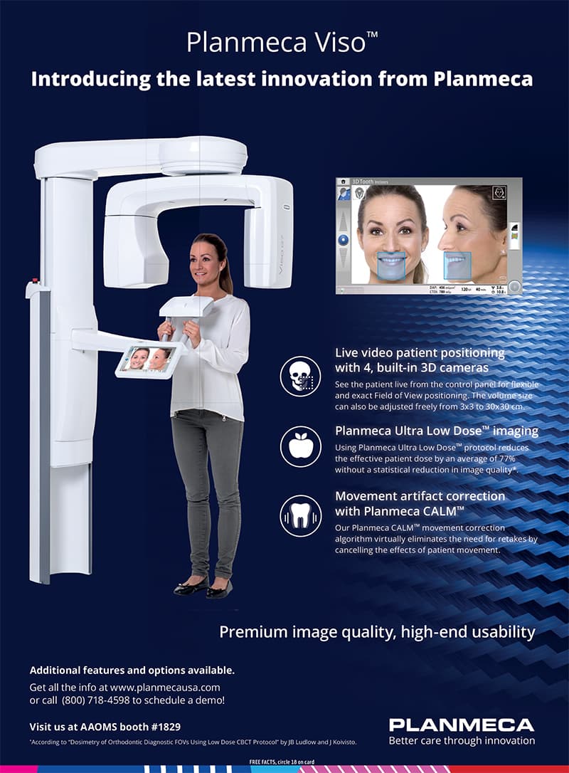
Cone-beam computed tomography, or CBCT, uses a rotating cone-shaped X-ray beam to reconstruct a three-dimensional volume. It can reveal selected dental and maxillofacial relationships that two-dimensional images may not show adequately, but it uses ionizing radiation and should not be a routine screening spectacle. Current ADA-AAOMR recommendations call for history, examination, disease-risk assessment, and review of prior images before imaging, with CBCT used judiciously when lower-exposure options will not provide the information needed. Planmeca documents that the Viso G7 platform offers adjustable fields up to 30 × 30 cm, 120 kV imaging, a 75 μm endodontic mode, Ultra Low Dose protocols, CALM motion correction, and optional ProFace facial capture. Those are manufacturer capabilities—not proof that every module is licensed, used, or appropriate for a particular patient.
For
01
A dental, endodontic, implant, surgical, orthodontic, or jaw-bone question for which three-dimensional information may change care.
02
Someone whose prior two-dimensional images do not adequately answer a defined anatomic or diagnostic question.
03
A patient who wants to understand why a scan is proposed, which area will be included, how exposure is limited, and who will interpret the complete volume.
01 — Ask the question
History, examination, disease risk, and available prior imaging establish what information is missing and whether another imaging method can answer it.
02 — Select and optimize
The field of view, resolution, and exposure settings are matched to the patient, anatomy, and diagnostic task so the needed image quality is obtained without unnecessary coverage or dose.
03 — Interpret the complete volume
The entire acquired volume is reviewed for relevant findings and limitations, with oral and maxillofacial radiology consultation when the scope or finding requires it.
04 — Connect image to care
The scan is considered with the clinical record and restorative or surgical objective; it does not independently determine a diagnosis, implant position, procedure, or outcome.
CBCT guide
The best scan is the one that answers the question with the least necessary exposure.
A large field and fine voxel are technical options, not measures of better care. The patient-specific benefit must outweigh the radiation risk, and every setting should serve a defined diagnostic or treatment-planning task.
When three dimensions may add value
CBCT may be considered for selected implant, impacted-tooth, endodontic, trauma, developmental, pathologic, orthodontic, or osseous jaw questions when clinical findings and lower-exposure imaging do not provide enough information. The indication should be recorded before the scan, not invented from the scan afterward.
Review prior imaging first
Recent diagnostic images may already answer the question or allow a smaller additional examination. Avoiding duplicate imaging is part of justification. Frequency is based on current findings and risk, not a fixed calendar or a premium-service package.
Field of view and resolution are selected
The field should cover the area of interest without unnecessary anatomy. Smaller voxels can show finer detail but may require different exposure and can increase noise or artifact sensitivity. The operator balances coverage, resolution, patient size, movement, metal, and the diagnostic task.
Radiation benefit and risk
FDA and ADA guidance supports justification and optimization: use CBCT only when needed, consider lower- or non-ionizing alternatives, discuss rationale and risk, and choose the lowest exposure that provides adequate diagnostic quality. Children and younger patients require particular care because radiation risk is more consequential over a longer lifetime.
What the manufacturer documents
For the Viso G7 platform, Planmeca documents fields up to 30 × 30 cm, 120 kV operation, 75 μm endodontic imaging, Ultra Low Dose protocols, CALM motion-artifact correction, and ProFace as an option. A maximum field is not the default, low-dose branding does not make an unjustified scan appropriate, and artifact correction cannot remove every limitation.
What CBCT does not show equally well
CBCT is especially useful for mineralized structures but has limitations in soft-tissue contrast. For TMD, for example, CBCT can address selected bone questions while MRI is preferred for the articular disc and other joint soft tissue. Motion, metal, reconstruction, field size, and acquisition choices can create artifacts or obscure detail.
Interpretation carries responsibility
The complete acquired volume should be reviewed, not only the tooth or implant site that prompted the scan. Incidental or uncertain findings may require comparison, follow-up, medical or dental referral, or interpretation by an oral and maxillofacial radiologist. An image is evidence within an examination—not a diagnosis by itself.
Practice verification before indexed publication
The clinic must match the named model to a current equipment record; confirm address, ownership, registration, inspection and quality-assurance status; document operator training and interpretation workflow; and list only licensed modules actually in use. No outcome, exclusivity, dose, accuracy, or safety claim should be inferred from the model name alone.
Continue the evaluation
Clinical reading
General information only. Diagnosis, treatment, risks, alternatives, and expected recovery are individual and require a clinical evaluation.
CBCT earns its place when a documented question requires three-dimensional information and the result can change care.
Request a visit