
Clinical technology
Every machine answers to the case.
Named systems, their proper place in the workflow, and the limits of what technology can claim.
01 / 3D imaging
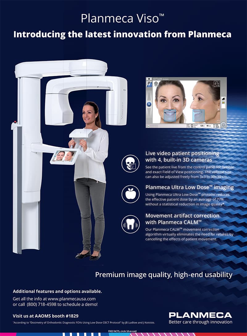
Planmeca Viso G7
An adjustable CBCT platform with fields of view up to 30 × 30 cm. Planmeca documents a 75 μm endodontic mode, 120 kV imaging, Ultra Low Dose protocols, CALM motion correction, and ProFace facial capture. The field and protocol are selected for the anatomy and diagnostic question—not for spectacle.
02 / Review and planning
Planmeca Romexis
The imaging volume can be reviewed in axial, coronal, sagittal, and case-specific planes. Relevant anatomy can be studied alongside other digital records to support diagnosis and surgical or restorative planning.
03 / Functional records
K7 · surface EMG · TENS
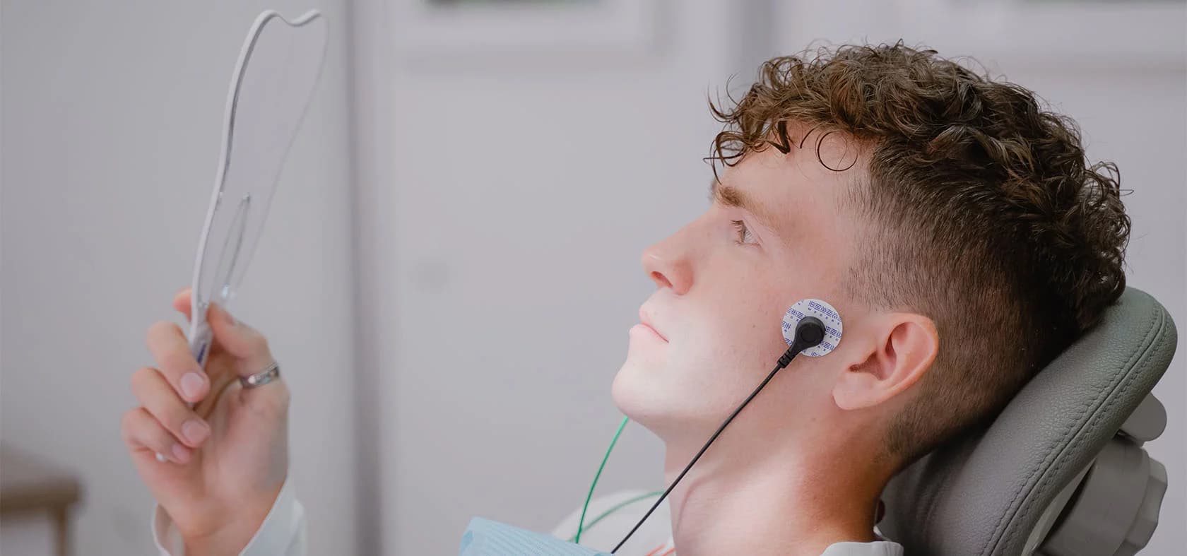
When clinically indicated, computerized mandibular scanning tracks jaw movement, surface electromyography records muscle activity, and TENS may be used during a functional evaluation. These records inform the examination; they do not replace a diagnosis or guarantee an outcome.
04 / Implant position capture
PIC photogrammetry
For selected complete-arch implant cases, photogrammetry captures the relative positions of implant components for the digital restorative workflow. Accuracy depends on the validated system, calibration, acquisition, and case conditions; no clinic-specific tolerance is claimed here until the installed model is documented.
05 / Design and fabrication
Digital laboratory
Clinical and laboratory teams coordinate the digital model, proposed bite, materials, design, and fabrication in the same building. The exact mill, materials, validated tolerances, and turnaround are being inventoried for publication and remain case-specific.
The coordinated process
Technology is valuable when the handoff disappears.
01
Ask the clinical question
The examination determines which record could change the plan.
02
Capture only what is useful
A focused or larger field is selected according to anatomy, diagnosis, and radiation principles.
03
Align the records
Imaging, photographs, scans, functional data, and history are reviewed as one case.
04
Test the plan
Provisional, orthotic, surgical, or restorative phases are chosen to make critical assumptions observable.
05
Design and fabricate
The laboratory works from the approved clinical position, material plan, and case-specific objectives.
06
Verify and maintain
Fit, function, tissue response, home care, and follow-up remain part of the result.
Verification standard
Model names, features, and performance figures will remain linked to manufacturer documentation and the practice’s installed-equipment inventory. Clinical outcomes will never be attributed to a machine alone.
Request a consultation →