11 — Work
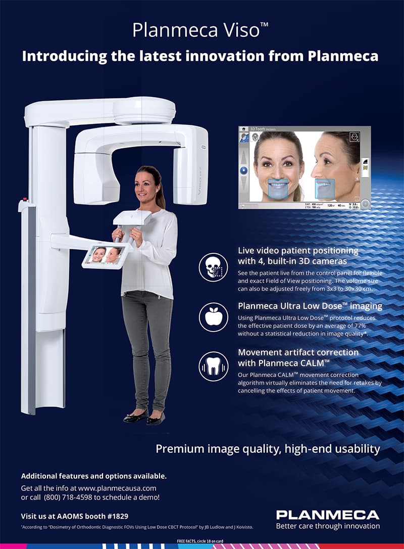
Planmeca Viso G7
The whole skull. The neck. The joint in motion. Then we operate.
What it is
This practice has installed a Planmeca Viso® G7 — the flagship CBCT in that line, with a freely adjustable field of view from about 3 × 3 cm (a single tooth at 75 μm) to 30 × 30 cm. That upper volume reaches from the skullcap through the face to C7 on the cervical spine. In Planmeca Romexis we do not look at a flat picture. We dissect the volume: axial, coronal, sagittal, and arbitrary planes; we segment airway, jaws, teeth, sinuses, and nerves; we sit with the patient in the reconstruction of their own head. 4D Jaw Motion can record the mandible moving in three dimensions — the joint as a living organ, not a still. ProFace captures a 3D photograph with four cameras in the sensor and composites it onto the bone, so the face that will wear the arch is on the same model as the implants. Ultra Low Dose and CALM (motion correction) keep the capture usable for a traveler who cannot sit for a retake. 120 kV reduces artefact around existing metal. This is not a gadget in the corridor. It is how a gneuromuscular reconstruction is allowed to start: the joint, the airway, the neck, and the bone in one model, then PIC, then the mill.
For
01
A joint that has never been seen in three dimensions, only guessed at from a panoramic.
02
A full-arch or failed-implant case that needs true bone, airway, and neck — not a cropped jaw.
03
A patient who needs to understand the plan: we can walk through their own skull, slice by slice.
01 — Capture the volume that the case actually needs
Live cameras on the unit set the field of view with the patient facing us. A tooth at 75 μm, both TMJs, the face, or a full skull to C7 — not a default box.
02 — Dissect, model, show
Romexis slices the volume. Airway and nerve can be segmented. The patient can see why a pretty arch on this bite, this neck, this joint would fail. 4D motion when the mandible must be watched, not imagined.
03 — Operate on a virtual patient that matches the person
CBCT + ProFace + intraoral / PIC coordinates. Implant positions in true bone. Vertical dimension against the airway and the cervical spine. Then surgery and the mill in the same building.
Most rooms still plan a face from two dimensions. We will not.
Request a visit